DFG - Bimodal reconstruktion and Magnetic Particle Imaging
| Working Group: | WG Industrial Mathematics |
| Leadership: | Prof. Dr. Dr. h.c. Peter Maaß ((0421) 218-63801, E-Mail: pmaass@math.uni-bremen.de ) |
| Processor: |
Christine Bathke
Dr. Tobias Kluth |
| Funding: | CDZ, DFG |
| Project partner: |
Prof. Ming Jiang, PhD, Peking University, China Prof. Dr. Alfred K. Louis, Universität des Saarlandes Prof. Dr. Christina Brandt, Universität Hamburg |
| Time period: | since 01.08.2015 |
For medical diagnosis there exists a variety of imaging methods, e.g. computed tomography (CT), magnetic resonance imaging (MRI) and positron emission tomography (PET). Common to all of these methods is their limitation to one specific physical quantity. Therefore, one method can only image information related to that particular quantity. It seems only natural to combine different imaging methods to get more information on the patient. In most cases functional imaging methods, that for example show metabolic processes or blood flow, are combined with methods that image anatomy.
Usually the images from different methods, also called modalities, are first reconstructed independently of each other and then overlayed using a registration method.
Bimodal reconstruction, however, combines the information of two modalities already during the reconstruction process, assuming that specific features, e.g. edges, are common to different modalities. Thus we do not only get information on the anatomy as well as on body functions, that complement each other, but we can can also profit from the higher resolution of the established anatomic tomography methods. Furthermore, bimodal reconstruction allows to supplement missing information using the second modality.
A promising method, that has gained popularity in recent years, is the so-called magnetic particle imaging (MPI). It is capable of imaging bloodflow as well as tracking instruments during a medical intervention by using a magnetic tracer. It does not need any harmful radiation, while it offers a high resolution for this kind of application. Since the sole reconstruction of the tracer concentration does not show its location inside the patient, magnetic particle imaging is often combined with magnetic resonance imaging. In this way the tracer can be located in specific blood vessels and organs.
To reconstruct the concentration of the MPI tracer while taking into account anatomical information from magnetic resonance imaging, we tested an extension of the total-variation functional (TV functional). This extension uses a projection of the edges from MPI to the the additional information and at the same time favors piecewise constant areas as the classic TV functional does.
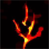
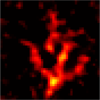
First simulations showed the potential benefit of this reconstruction approach for magnetic particle imaging. Compared to the Kaczmarz method, that is most commonly used for magnetic particle imaging so far, the reconstruction with additional information delivers significantly better results with sharper edges and a background that is more homogenuous (see figures below). The quality of the reconstruction is especially improved at the boundaries of the measurement area, where the signal quality of magnetic particle imaging is considerably worse than in the center due to the physics of the MPI scanner.
Here, the additional anatomical information obtained by MRI can partially substitute the missing information on the tracer distribution from the MPI scan due to many common edges.
Artery 1 to 3
left: simple reconstruction with noisy/incomplete data - blurred edges and noticeable background noise
center: additional information from a second modality - partly matching the image to be reconstructed
right: reconstruction with additional information - sharper edges and smoother background

