
DIAMANT - Digital Image Analysis and Imaging Mass Spectrometry to Differentiate Non-small Cell Lung Cancer
| Working Group: | WG Industrial Mathematics |
| Leadership: | Prof. Dr. Dr. h.c. Peter Maaß ((0421) 218-63801, E-Mail: pmaass@math.uni-bremen.de ) |
| Processor: |
Dr. Charlotte Janßen
Dr. Daniel Otero Baguer Dr. Lena Hauberg-Lotte |
| Funding: | BMBF Computational Life Sciences |
| Project partner: |
Prof. Dr. Jörg Kriegsmann, MVZ für Histologie, Zytologie und Molekulare Diagnostik Trier Dr. Katharina Kriegsmann, Universitätsklinikum Heidelberg |
| Time period: | 01.01.2020 - 31.12.2022 |
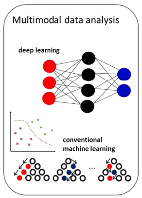
Despite all the progress made in the development of new therapeutic methods, cancer remains one of the most serious health threats and most frequent causes of death worldwide. For optimal treatment of patients, early and accurate histopathological diagnosis of tumors and their respective subtypes is of utmost importance. Although some diagnostic tools are available for this purpose in clinical routine, in some cases a considerable degree of uncertainty remains. The DIAMANT project focuses on lung cancer, the most common cancer in men worldwide and the second most common cancer in women. The main subtypes are adenocarcinoma (ADC) and squamous cell carcinoma (SqCC), which together account for ~70% of all lung cancers. The distinction between these subtypes is very important, as some therapies are promising for one type and contraindicated for another. In poorly differentiated tumours, however, typing is often not possible based on morphology alone, but requires additional immunohistochemical (IHC) staining with up to four markers. In these cases, less extensive processing leads to reduced diagnostic accuracy, with the risk of delayed or even incorrect diagnosis. To solve this problem in the context of tumor classification, the use of matrix-assisted laser desorption/ionization mass spectrometry (MALDI IMS) has been proposed. MALDI IMS is an analytical method for the spatially resolved examination of proteins, metabolites, lipids and other macromolecules in biological tissue samples. A major advantage is its applicability to formalin-fixed, paraffin-embedded tissue sections, as they are predominantly used in the histopathological routine. In recent years, promising results have been obtained regarding the MALDI IMS based classification of different cancer types. However, the method has not yet been established in clinical routine. One reason for this is the limited spatial resolution of MALDI IMS (approx. 50 μm), which does not allow molecular imaging at cell level. The DIAMANT project therefore combines the molecular information from MALDI IMS with the detailed anatomical information from digital microscopy images (Digital Image Analysis, DIA). By means of an integrated analysis of the data from both complementary modalities, a classification model will be developed that is significantly more accurate than existing models based on only one of the two modalities. The deep learning methodology used in the project is particularly well suited to extract meaningful information from the complex, high-dimensional raw data of both modalities. Using an application-specific network architecture and specially adapted regularization methods, pathological, biochemical and mass spectrometric a-priori knowledge is integrated into the classification model. This reduces the required amount of training data and increases the robustness of the method and its generalizability to new clinical test data.